

An OPG scan is one wide X-ray of all your teeth and both jaws. Your dentist may have just referred you for an OPG scan. So you probably wonder why, how it feels and what the dose is. This guide answers each question in plain English.
What Is an OPG Scan?
OPG stands for orthopantomogram. It is the image an X-ray arm records as it circles your head. Unlike small films placed inside the mouth, it covers the whole lower face. So your dentist sees every tooth and the surrounding bone in one flat view.
Why the name sounds so complicated
The full name describes a curved slice through both jaws, recorded in one sweep. A referral may mention a panoramic scan called an OPG, a DPT or a “pan”. Whatever the label, an OPG scan is the same short examination.
- OPG (orthopantomogram)
- A panoramic X-ray taken from outside the mouth that shows all the teeth and both jaws in one image.
- Bitewing
- A small X-ray held between the back teeth to show decay between them and around fillings.
- Periapical
- An intraoral X-ray that shows one or two teeth in full, down to the root tips.
- CBCT (cone beam CT)
- A 3D dental scan that measures bone and nerve position, with a higher dose than an OPG.
- Microsievert (µSv)
- A unit of effective dose; 1,000 microsieverts equal one millisievert.
Why Would a Dentist Ask for One?
An OPG scan shows buried and unerupted teeth and the height of the jawbone. It covers the jaw joint area and the floor of the sinuses, too. Cysts and other changes inside the bone may also appear. That is why dentists often request one at a first visit or before surgery.
Wisdom teeth and buried canines
Lower wisdom teeth sit close to the nerve that supplies the lip and chin. Before any wisdom tooth extraction, the OPG dental scan shows the tooth’s angle and roots. It also shows how near those roots come to the nerve canal. Dentists map buried canines the same way before impacted tooth extraction.
Implants and orthodontics
For a single tooth implant, the panoramic image gives a first look at the bone. Precise measurements, however, usually need a 3D scan afterwards. In children, orthodontists use it to follow which adult teeth are coming through.
Where an OPG Scan Falls Short
An OPG scan squeezes a curved jaw into one flat picture. Because of that, it trades fine detail for breadth. Small cavities between teeth and early root-tip changes can hide in the overlap. The picture has slight magnification and soft areas at the front. So it is not a tool for exact measurements on its own.
Bitewings for decay between teeth
A bitewing sits inside the mouth and shows the back teeth up close. As a result, it spots early decay between teeth far better. When one root needs a closer look, a periapical film does the job instead. Both take seconds. So many check-ups pair an OPG scan with one or two of them.
OPG, Bitewing or CBCT: Which Image Does What?
Think of the OPG as a map and CBCT as a 3D model. The map shows the whole area at a modest dose. Cone beam CT, by contrast, measures bone width and nerve position in three dimensions. Each answers a different question, so dentists often combine them.
| Image | What it covers | Typical use | Effective dose |
|---|---|---|---|
| OPG (digital sensor) | All teeth and both jaws, 2D | Wisdom teeth, bone level, first assessment | 14.2–30 µSv |
| Bitewing (single, digital) | Crowns of the back teeth | Decay between teeth, edges of fillings | 0.3–1.4 µSv |
| Bitewings (set of four) | Back teeth on both sides | Routine decay checks | 3.4–5 µSv |
| CBCT, small field (adult) | Chosen area, 3D | Implant planning, tooth close to the nerve | 19–652 µSv |
| CBCT, large field (adult) | Most of the face, 3D | Complex surgical planning | 68–1,073 µSv |
Doses come from Table 1 of the ADA panel report (2024) and vary by machine.
When a 3D scan is worth the extra dose
CBCT earns its place when an implant needs millimetre planning. It helps, too, when a buried tooth sits right against the nerve. The ADA panel advises against routine CBCT and keeps it for questions lower-dose images cannot answer. In short, an OPG scan usually comes first and CBCT follows only if needed.
Keeping the field small
The same report says CBCT can reach 10–15 times the dose of standard dental X-rays. Dose rises with the size of the area scanned. So clinicians choose the smallest field that covers the region in question.
What Happens During the Appointment?
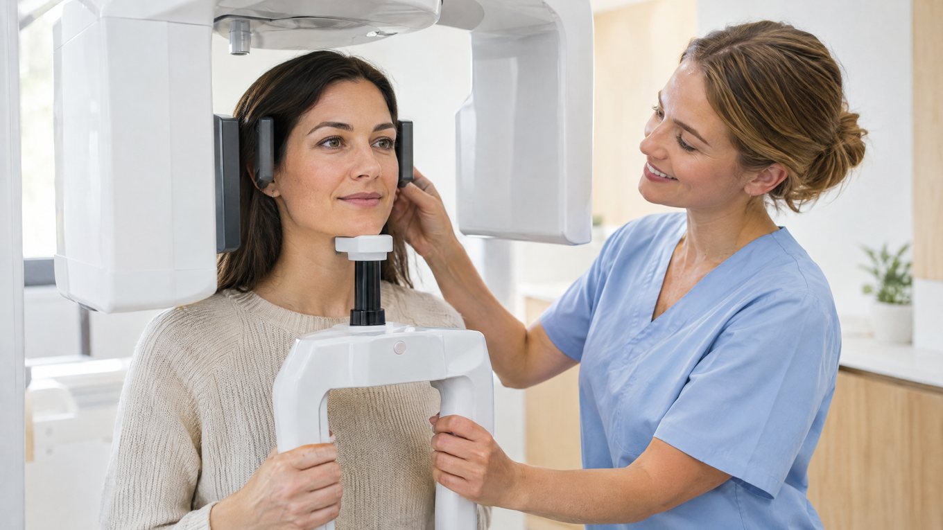
The scan is painless and needs no preparation. You stand at the machine and bite gently on a plastic guide. Then you keep still for 12 to 20 seconds as the arm circles your head. From start to finish, an OPG scan usually takes only a few minutes. After that, digital images appear on screen straight away.
- First, remove earrings, necklaces, glasses, hair clips and any removable denture or retainer.
- Mention any chance of pregnancy before the scan starts.
- Step up to the machine and rest your chin on the support.
- Bite lightly on the guide with your front teeth.
- Press your tongue to the roof of your mouth and close your lips.
- Stay still until the arm has finished its sweep.
Why the jewellery has to come off
Metal blocks X-rays, so earrings leave bright shadows over the jaw. A shadow in the wrong place may mean a repeat scan and extra dose. Fixed fillings, crowns, implants and braces stay in, because your dentist can read around them.
How Much Radiation Does an OPG Scan Involve?
Very little, although it is not zero. The ADA’s 2024 expert panel lists 14.2 to 30 microsieverts for a digital panoramic image. Its comparison uses the US average of 3.1 millisieverts a year. On that basis, the dose matches roughly 2–3.5 days of natural radiation. Even so, each OPG scan should answer a real clinical question.
Digital (CCD) panoramic X-ray: 14.2–30.0 µSv, about 2–3.5 days of background radiation. Small-field adult CBCT: 19–652 µSv, about 2–77 days.
Benavides E et al., Journal of the American Dental Association 2024;155(4):280–293, Table 1.
Will I wear a lead apron?
Yes. At our clinic, you wear a protective lead apron during the scan. Some recent guidelines no longer consider routine shielding mandatory. For instance, the ADA’s 2024 expert panel and the FDI World Dental Federation take this view. Practice still varies between countries, so feel free to ask our team about protection.
Pregnant or Trying for a Baby? Tell Us First
An OPG scan targets the jaws, far from the womb, at a low dose. Even so, tell the team before the scan if you are or might be pregnant. Non-urgent images can usually wait until after the birth. But an X-ray your treatment truly depends on should not be withheld. The FDI World Dental Federation makes the same point.
Bring your previous X-rays
If another dentist took a panoramic image recently, bring a digital copy. A recent image may then make a new OPG scan unnecessary. The ADA asks dentists to make a good-faith effort to obtain earlier radiographs. Our guide to dental care during pregnancy answers other common questions. Travelling to Istanbul for treatment? Send recent images ahead of your visit.
Before your appointment
Is an OPG scan the same as a normal dental X-ray?
Can I eat and drink normally before an OPG?
Will I feel claustrophobic in the machine?
Can I keep my brace or crowns in?
Safety, frequency and results
How often can I safely have a panoramic X-ray?
Do children have OPG scans?
Does a CBCT scan involve more radiation than an OPG?
How quickly will I see the image?
Now you know what an OPG scan shows and where it falls short. Want to know whether you need one? Contact our clinic or meet our dentists before your visit. Our treatments page lists every service we offer. More patient guides wait on the Smile Group blog.
Written by: Smile Group Clinical Team · Reviewed by our dentists · Last updated: 20 October 2026
Sources: Benavides et al., JADA (2024) · ADA — X-rays/Radiographs · ADA — 2024 radiography recommendations · FDI — Radiation safety in dentistry · RadiologyInfo — Panoramic dental X-ray